
Mutations that change protein structureSpec D1.2.11
In short
A mutation that changes the base sequence of a gene can change the amino acid sequence of its polypeptide, and so the protein's structure. In sickle-cell anaemia, a base substitution in the haemoglobin beta-chain gene changes the codon GAG to GUG, so valine replaces glutamic acid. Haemoglobin S forms fibres at low oxygen, distorting red blood cells.
The amino acid sequence of a polypeptide decides how it folds and therefore its three-dimensional structure. A change in the base sequence of a gene can change the codons in the mRNA, and so the amino acid sequence, the folding and the function of the protein.
Example: sickle-cell anaemia
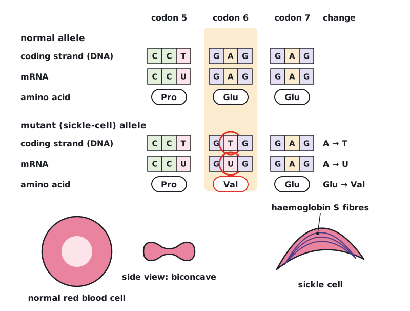
- The gene for the beta chain of haemoglobin has a base substitution: on the coding strand, A is replaced by T (GAG becomes GTG).
- The mRNA codon becomes GUG instead of GAG.
- At the sixth amino acid of the beta chain, valine replaces glutamic acid. The resulting form is haemoglobin S.
- Glutamic acid is charged and hydrophilic; valine is non-polar and hydrophobic. At low oxygen concentrations, haemoglobin S molecules stick together into long fibres.
- The fibres distort red blood cells into a rigid sickle shape. Sickle cells can block capillaries and carry less oxygen, and they are broken down faster, causing anaemia.
A change to just one base of the gene is enough to alter the protein. This shows how closely protein structure depends on base sequence.
 (opens full size in a new tab)
In a sickle-cell answer, give the base change, the codon change (GAG → GUG), the amino acid change (Glu → Val) and the effect on haemoglobin and red blood cells. Each is a separate marking point.
Written and checked against the IB Biology HL specification · Updated October 2026