Sliding filament model of muscle contractionSpec B3.3.2
In short
The sliding filament model explains muscle contraction: myosin heads bind to actin filaments, forming cross-bridges, and pull the actin towards the centre of the sarcomere using energy from ATP. The actin and myosin filaments slide past each other without changing length, so the sarcomere, and the whole muscle, shortens.
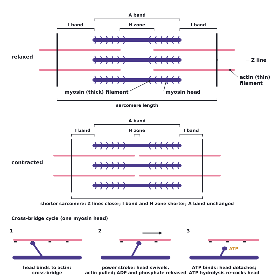
A skeletal muscle fibre contains many myofibrils. Each myofibril is a chain of repeating units called sarcomeres, separated by Z lines.
- Thin actin filaments are attached to the Z lines and point towards the centre of the sarcomere.
- Thick myosin filaments lie in the centre, overlapping the actin. Each has many myosin heads.
- The dark band (A band) is the length of the myosin; the light bands (I bands) contain only actin; the H zone in the centre contains only myosin.
- A nerve impulse causes calcium ions to be released from the sarcoplasmic reticulum. They bind to regulatory proteins on the actin, exposing binding sites for myosin.
- Myosin heads, already energised by hydrolysing ATP, bind to the binding sites on actin, forming cross-bridges.
- The heads swivel (the power stroke), pulling the actin filaments towards the centre of the sarcomere. ADP and phosphate are released.
- A new ATP binds to each myosin head, which then detaches from the actin.
- Hydrolysis of ATP re-cocks the head, ready to bind further along the actin. The cycle repeats while calcium is present.
The filaments slide past each other; neither changes length. During contraction the Z lines move closer together, the I band and H zone shorten, and the A band stays the same length.
 (opens full size in a new tab)
The filaments do not shorten or contract. They slide past each other, so only the sarcomere shortens.
Written and checked against the IB Biology HL specification · Updated October 2026