Translation at the ribosomeSpec D1.2.5, D1.2.6, D1.2.7, D1.2.10
In short
Translation is the synthesis of polypeptides from mRNA: the base sequence of mRNA is translated into the amino acid sequence of a polypeptide. mRNA binds to the small ribosomal subunit, tRNA anticodons pair with mRNA codons, and the ribosome moves along the mRNA one codon at a time, linking amino acids by peptide bonds to the growing chain.
Translation is the synthesis of polypeptides from mRNA. The base sequence of the mRNA is translated into the amino acid sequence of a polypeptide. It takes place at ribosomes in the cytoplasm.
| Molecule | Role |
|---|---|
| mRNA | Carries the base sequence copied from a gene. It binds to the small subunit of the ribosome. Its bases are read in groups of three (codons). |
| Ribosome | Made of a small and a large subunit (rRNA and protein). Two tRNAs can bind simultaneously to the large subunit, so their amino acids are held next to each other. The ribosome catalyses peptide bond formation. |
| tRNA | Each tRNA carries a specific amino acid and has an anticodon of three bases that pairs with a matching codon on the mRNA. |
Codons and anticodons
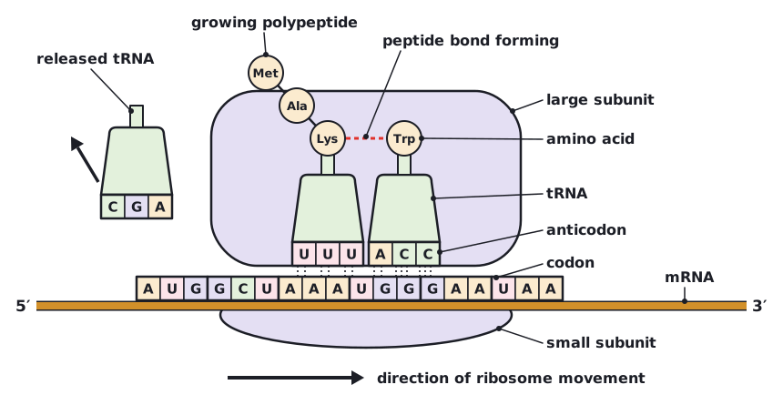
A codon is a sequence of three bases on mRNA that codes for one amino acid. An anticodon is the complementary sequence of three bases on a tRNA. The anticodon pairs with the codon by complementary base pairing and hydrogen bonding (A with U, C with G), which ensures the correct amino acid is added. For example, the codon AUG pairs with the anticodon UAC.
Elongation of the polypeptide
- A tRNA carrying the growing polypeptide is bound to the ribosome, with its anticodon paired to a codon on the mRNA.
- A second tRNA, whose anticodon is complementary to the next codon, binds alongside it, bringing its amino acid.
- A peptide bond forms between the amino acid on the second tRNA and the polypeptide, so the polypeptide is transferred to the second tRNA and becomes one amino acid longer.
- The ribosome moves along the mRNA by one codon (three bases). The first tRNA, now without an amino acid, is released and can collect another amino acid.
- The next codon is exposed and the cycle repeats, adding one amino acid at a time until a stop codon is reached.
This stepwise movement of the ribosome along the mRNA, one codon at a time, means the amino acids are linked in exactly the order set by the codons.
 (opens full size in a new tab)
Codons are on mRNA and anticodons are on tRNA. Do not describe the anticodon as part of the mRNA or the codon as part of the tRNA.
Written and checked against the IB Biology HL specification · Updated October 2026